There are two basic types of microscopes: The older type with objectives corrected for 160 mm tube length and the modern type with infinity-corrected objectives (details here). The infinity construction has the advantage that various auxiliary components (compensators, filters, epifluorescence mirrors, etc.) can be inserted into the ray path without changing the optical configuration of the entire system. This is an enormous simplification in microscope design. In order to focus the image of the specimen, a secondary lens, the the tube lens is necessary to project a visible picture into the eyepiece. So far for microscopes.
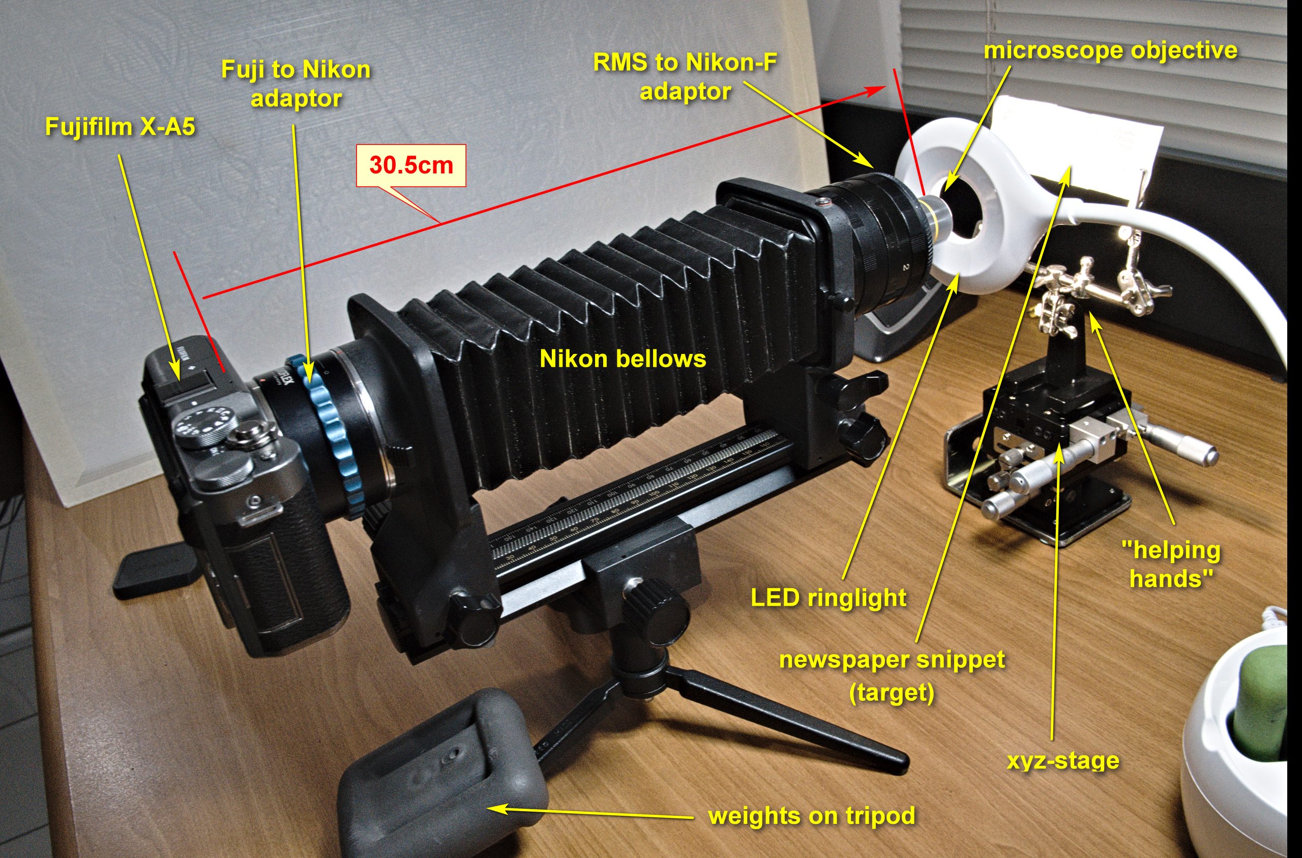
In the recent years, microscope objectives are being used directly on a camera (as shown in Figure 1) for extreme macro photography with reasonable success. However, a vigorous debate has ensued, if infinity-corrected objectives can be used in a conventional macro set-up without a tube lens. It is being argued (the internet is full of it) that such objectives cannot produce an image on the camera’s sensor because »they focus on infinity« (whatever that may mean). Practice has shown that infinity objectives can very well produce an image (as demonstrated in Figures 2 and 4, below). The remaining dispute focuses on image quality: Are images without a tube lens indeed inferior to those taken with the intermediate (ie. tube-) lens? This article tries to shed a new light onto the situation.
Another important aspect is the cost of microscope objectives: Second-hand 160mm objectives, now rarely used anymore, from an unbranded manufacturer in the Far East can be had for less than $50.-. In contrast, good branded infinity objectives sell for 10 to 20 times that prices and the prices for factory-new specialty objectives (fluorite, apochromatic, etc.) reach into the upper 4-digit dollar range. This begs the question: Is the image quality dramatically better to justify the cost? (Again: We are talking as macro objective on an (amateur-)camera, and not about the use on a modern microscope).
The Experiment
In this experiment, I am comparing two microscope objectives, both used without an intermediate (tube-)lens, which may, as some say, necessary when using infinity corrected objectives on a camera (as opposed to the use on a microscope). If a tube lens would be important in this configuration, we would expect a significant difference in image quality between the two objectives.
For the purpose of this comparison, we are using two fairly typical microscope objectives:
| Olympus | No-Brand (Chinese) | |
| Type | AchN-P (strain-free achromat) | achromat |
| Geometry | infinity corrected | Corrected for 160 mm tube length |
| Magnification | 10x | 10x |
| Numerical Aperture, NA | 0.25 | 0.25 |
The Set-up
For the comparison of the two objectives, we photographed a piece of regular newspaper, capturing part of a (black) printed character (See Figures 3 and 4). The »paper« was chosen to demonstrate planarity (flatness) of the field and the absence of substantial spherical aberration. Moreover, the black-and white scene of a newspaper print was expected to show chromatic aberration, which would manifest itself as green or purple fringing. which would be difficult to observe on colorful motives.
In addition, we photographed a customary microscope scale (glass-) slide to document the scale of magnification and as an example of a high-contrast motive. (See Figures 4 and 5).
Hardware
- Camera used: Fujifilm X-A5
- Sensor size: 23,5 x15.7mm, 6,000 x 4,000 pixels,
- Pixel size: 3.9 μm
- Extension: 30.5 cm (from the front of the objective to the sensor mark on the camera, see Figure 1), using a combination of Nikon bellows and Nikon extension tubes.
Processing and Software
- The series of images was recorded in RAW format (Fujifilm RAF format), files size 41,660 KB to be processed.
- Number of images per stack: 20
- Step interval: 3 μm (manual stepping)
The images were consequently converted to the JPG file format (for stacking) using a free software package named Rawtherapy. The file size of the JPGs was around 25,000KB (at 100% JPG-quality). During the RAW→JPG conversion process, only exposure, white-balance and contrast correction was applied. No dust mask, black frame (to remove hot pixels) or other quality-shaping steps were taken. It is important to mention that no sharpening or similar process was applied at this or any other stage of the workflow.
Stacking
The JPGs were stacked using the Helicon Focus software package. All of the several stacking algorithms offered in this package produced reasonable and useful results. The pictures shown in this context were all processed with Helicon’s algorithm ‘B’, »depth map«, subjectively the best results by visual comparison.
Comparison
All images in this article are show the uncropped, full-frame of the image, unless stated otherwise in the image caption. The images are also linked to the original image, which can be viewed and downloaded at full magnification for detailed viewing and scrutiny.

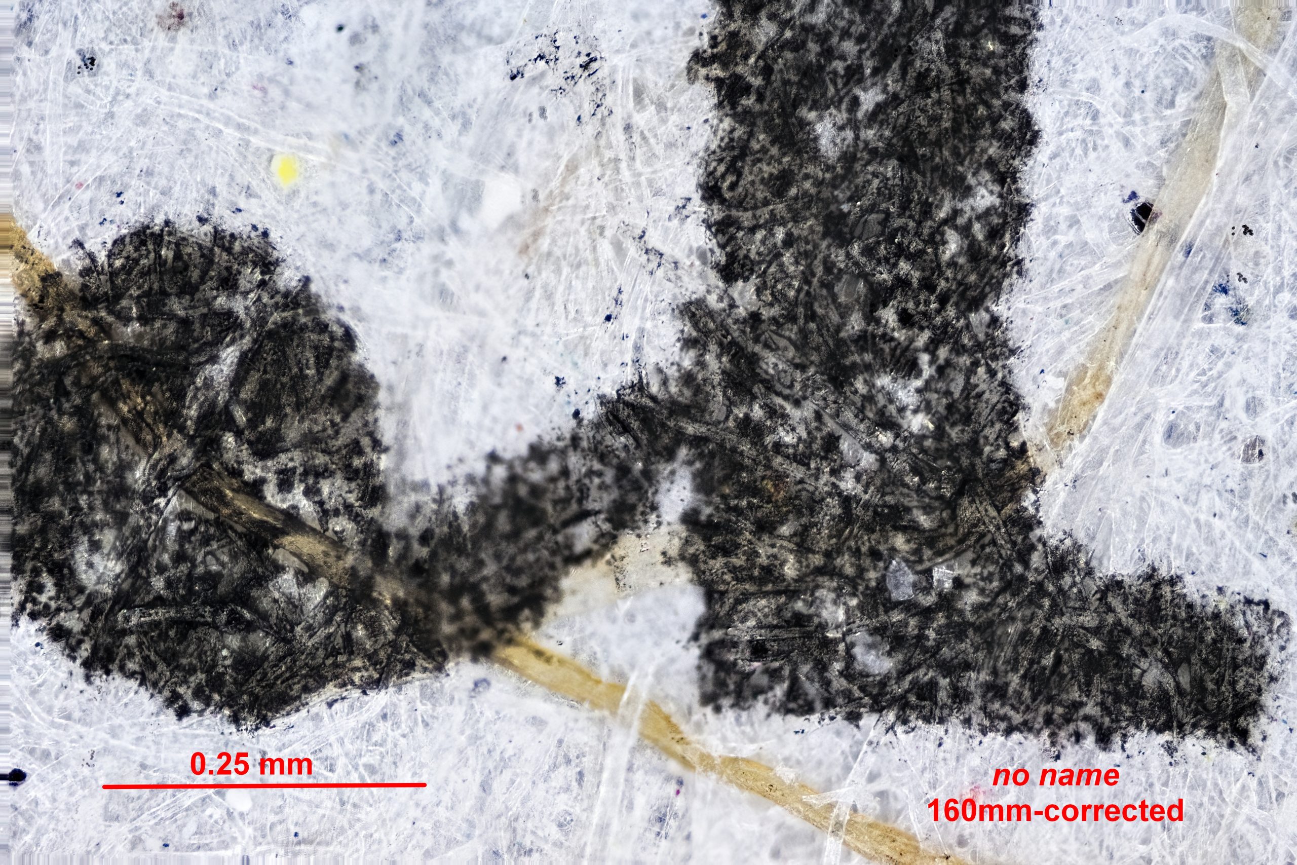
The Figures 2 and 3 show a similar newspaper snippet photographed in white light (4,900ºK). Visual inspection does not reveal major differences in the image quality. Moreover, chromatic aberration, manifested by color seams may exist but is not conspicuous, can be neglected. Also, there is no deterioration of the image quality towards the margin and corners of the images.

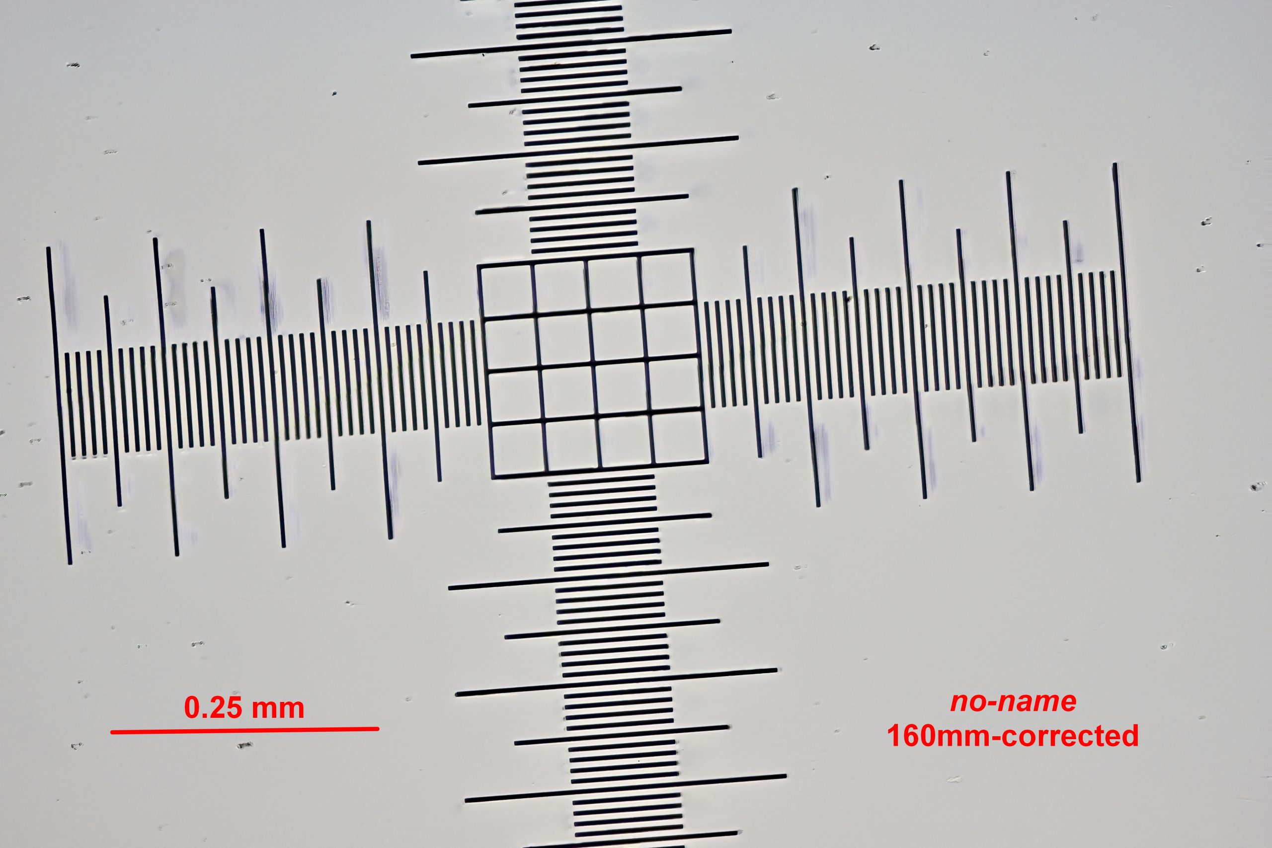
The Figures 4 and 5 show a microscope scale slide photographed with an illumination from behind the slide.
The »ghost image«, the duplicate »shadow« lines (seen on Figure 5, left side) have nothing to do with the optical quality of the objective. These shadows are artifacts from reflections within the the glass body of the microscope slide. (The light source was not in an optimal position).
Discussion of Results
Interestingly, the magnification of both 10x objectives is not the same, when they are used in the shown configuration on a camera. The magnification of no-name 160mm objective is about 30% (linear) larger.
The theoretical maximal resolving power of a 10x plan-apochromatic objective is – depending on wavelength – about 1.10 μm. This relates to roughly to 1/3 of a single pixel (of 3.9 μm) on the camera’s sensor. Close visual scrutiny of the presented images at 1:1 shows that details of at 4-6 pixels can be differentiated ‒ across the field and with both objectives. ‒ See for yourself ‒ you can download all images in original size and quality ‒ and, please, make your own conclusions!
Conclusion
Despite the substantially different design and manufacturing properties of the two objectives and against the backdrop of expectations, no substantial quality differences can be observed between the two types of objectives with the given set-up. However, when approaching the theoretical resolving power of the system, quality differences are expected to become visible.
Both types of objectives produce useful images of reasonable quality when mounted directly in front of the camera (Figure 1).
- No chromatic aberration can be observed.
- The theoretical resolution of both objectives has not yet been reached (see previous paragraph).
- The scale of magnification is different, owing to the different construction and correction. Images from the 160 mm objective are about 1/3 larger than those from the infinity objective.
- The myth that infinity-corrected objectives need a tube lens, when used on a camera in order to produce useful images has been dispelled with this experiment. (But not so, when used on a microscope!)
The mention or use of a certain software, hardware, brands etc. does not constitute a recommendation in any way. Proprietary names in the text were only used in order to document and describe the work flow.